C.LIVE Caspase-3
Green NucView®
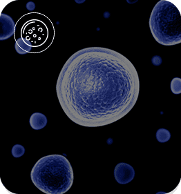
The C.LIVE Caspase-3 assay is a standardized, fluorescence-based assay that measures caspase-3 protease activity for apoptosis detection. This reagent contains a cell-permeable dye that is coupled to the caspase-3 substrate peptide sequence DEVD blocking its binding to DNA. If activated in apoptotic cells, caspase-3 cleaves the peptide sequence, and the dye is released to bind to the DNA with high-affinity, resulting in a fluorescent signal in the cell nucleus.
This allows for early detection of cell death independent of morphology changes of the cells. With one vial, you can analyze 400 samples (with 100 µL per sample) which is equal to 4 full 96-well plates of experiments. Together with the power
of the CELLCYTE X and C.LIVE reagents, open
the door to live cell imaging and view the
biological events in your cell samples with ease
Request Pricing
Access Product Brochure
![]()
Join 1,000+ Biopharmaceutical Companies and Academic Institutions
![]()
![]()
![]()
![]()
![]()
![]()
![]()

![]()
![]()

![]()


![]()
![]()

![]()
![]()
![]()
Key Features & Benefits
Find out how our product can enrich your research
Direct detection of activation of
caspase-3 as early indicator of
programmed cell death
Experience real-time detection of apoptosis inside the incubator.
Mix-and-read workflow
Seed, treat, and image your cells in three easy steps.
Optimized for live cell imaging analysis
Observe cell health behavior instantly without hinderance from optimal assay conditions.
Cell health integrity upheld
Confidently analyze your samples with the assurance that our reagent does not impair cell growth.
Product Details
Discover what you can doDirect, Real-time Detection of
Apoptosis Inside the Incubator
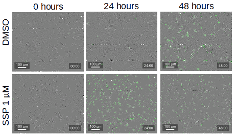
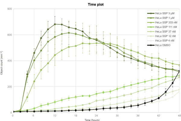
In the figure below, HeLa cells were treated with increasing concentrations of Staurosporine. C.LIVE Caspase-3 was added to each well (2.5 µM) and timelapse images were obtained with the CELLCYTE X. The DMSO control well shows minimal green signal compared to the Staurosporine-treated well at 48 hours (top panel). Automated quantification shows the time- and dose-dependent cytotoxic effects of Staurosporine (bottom panel).
Confidence in your cell
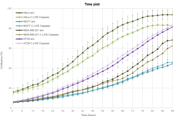
health integrityAs shown below, our caspase reagent does not impair cell growth, which gives you the assurance that your cell health integrity will remain upheld during the experiment. Whether you’re analyzing cancer cell health, drug screening, immune cell killing, or early toxicity, the combination of C.LIVE Caspase and the CELLCYTE X can handle any application.
Product
Applications
Accelerate your research
Apoptosis
Immune cell health
Immune cell growth
Immune cell differentiation
Immuno-oncology
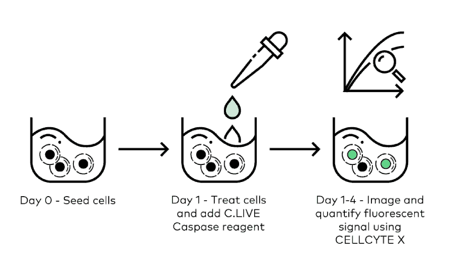
Featured Workflow
Run your apoptosis assays in three simple steps shown below.
Download Application Notes
For scientists by scientists, these notes highlight novel ways to optimize
your research with our products and solutions
Lorem ipsum Dolor sit
Lorem ipsum dolor sit
amet, consectetur
Lorem ipsum Dolor sit
Lorem ipsum dolor sit
amet, consectetur
Lorem ipsum Dolor sit
Lorem ipsum dolor sit
amet, consectetur
Product Datasheet
Donec scelerisque scelerisque neque, non sagittis ligula malesuada at.
| Product Name | Price | Channel | Amount | Concentration | Samples | Storage | Stability |
|---|---|---|---|---|---|---|---|
| C.LIVE Caspase-3 Green NucView® | $340 | Green | 20 µL | 5 mM (2000X stock solution) | 400 | -20°C | 12 months |
Lorem Ipsum is simply dummy text of the printing and typesetting industry. Lorem Ipsum has been the industry’s standard dummy text ever since the 1500s, when an unknown printer took a galley of type and scrambled it to make a type specimen book. It has survived not only five centuries, but also the leap into electronic typesetting, remaining essentially unchanged. It was popularised in the 1960s with the release of Letraset sheets containing Lorem Ipsum passages, and more recently with desktop publishing software like Aldus PageMaker including versions of Lorem Ipsum.
Lorem Ipsum is simply dummy text of the printing and typesetting industry. Lorem Ipsum has been the industry’s standard dummy text ever since the 1500s, when an unknown printer took a galley of type and scrambled it to make a type specimen book. It has survived not only five centuries, but also the leap into electronic typesetting, remaining essentially unchanged. It was popularised in the 1960s with the release of Letraset sheets containing Lorem Ipsum passages, and more recently with desktop publishing software like Aldus PageMaker including versions of Lorem Ipsum.
Featured Resources
Lorem Ipsum is simply dummy text of the printing and typesetting industry.


Previous
Next
Applications
Learn how you can automate research workflows in numerous application areas

Research

therapy

omics

isolation
development

microbial system
Previous
Next
See All Applications