

















Cellcyte X

Live-Cell Imaging and Analysis

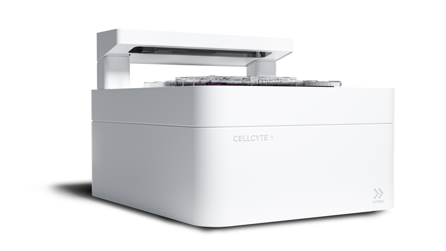
We recognize that science evolves with time and your research should as well. To leverage the power of continuity and address the most-pressing challenges in cell biology, we have developed the Cellcyte X, a high-throughput live cell imaging system that’s user-friendly, efficient, and robust.

  • GSK_logo_2022.svg
  • 211125_SANOFI_LOGO_RGB-1024x472
  • Amgen_logo_logotype-1-1024x239
  • novo-nordisk-1-logo-png-transparent-1-1024x1024
  • Bayer-Logo-1-1024x576
  • TevaPharm.svg-1-1024x359
  • Stanford_wordmark_2012.svg-1-1024x215
  • gilead-new
  • mit-logo-2-1
  • Pfizer-1-1024x419
  • Bristol-Myers_Squibb_logo_2020.svg-1-1024x165
  • 1200px-CSL_Limited_logo.svg-1024x516
  • johnson-johnson-logo-1-1024x185
  • Merck-1-1024x330
  • astrazeneca-logo-1-1024x248
  • Novartis-Logo.wine_-1-1024x683
  • Biogen_logo_logotype_symbol-1024x343
  • takeda_logo
  • Harvard_University_logo.svg-1-1024x268
  • LONZA_Logo
Cellcyte-Hero
BENEFITS

Bringing High-Throughput Live Cell Imaging to Your Lab

With our live cell imaging system hosted within the incubator, researchers can rewind and replay images acquired from multiple time points to better follow the sequence of biological events and get a comprehensive picture of cell kinetics. Read more below to see how the Cellcyte X can make a difference in your lab & analyses.

Open Design

Ease of maintenance and improved control of cellular environment.

Improved cell viability

Less disturbances over the course of your experiment, reducing the chances of cellular abnormality.

Real-time data analysis

With data collected and processed in real time throughout the experiment.

High throughput

Run 6 vessels concurrently to maximize your throughput.

Versatility

Multiplex your experiment with Enhanced Contour imaging mode plus 3 fluorescent channels.

Improved data output

Acquire and store thousands of images per experiment.

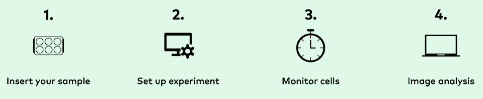
Simple 4-Step Workflow

Optimize your workflows while maximizing efficiency, saving time, and yielding highly reproducible data.

With an easy setup, the ability to monitor cell behavior in real time, intuitive software, and instant data visualization, you’ll have the tools you need for ultimate success in any application area. 

Screenshot 2026-07-02 at 8.57.55 PM
FEATURES

Built for Successful Experiments

Intuitive and comprehensive CELLCYTE STUDIO software
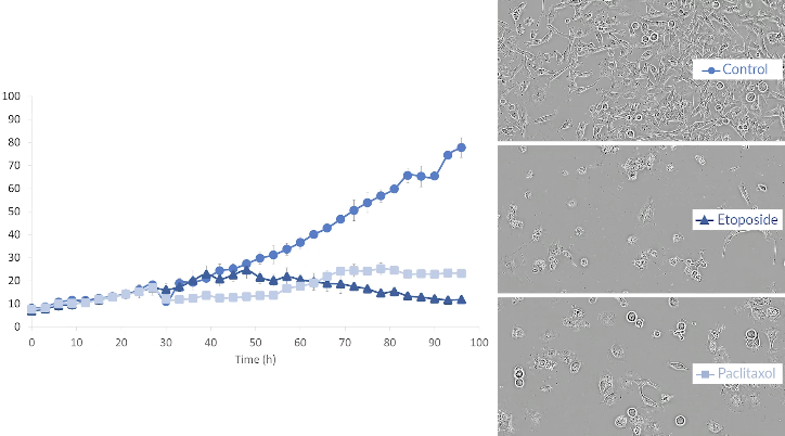
  • Analyze spheroids
  • Capture images and movies in real-time
  • Flexible scheduling options
  • Automated graphing tools
  • Platemap feature to plan out your studies ahead of time
  • 4x imaging
CELLCYTE-X-Real-time-analysis

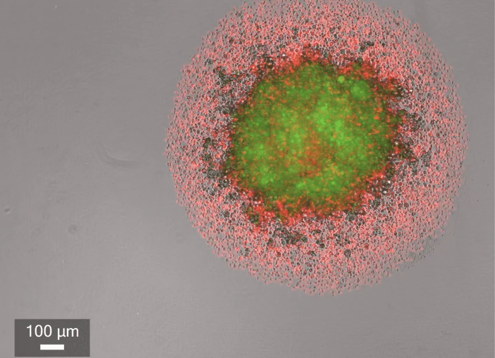
Real-time analysis of spheroid assays
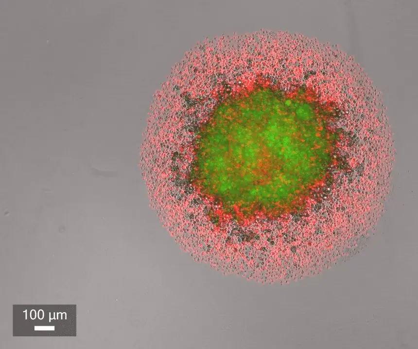
  • Streamlined workflow to monitor spheroid formations inside your incubator
  • Ensured autofocus at every timepoint for long-term live cell studies
  • Powerful analysis algorithm for accurate spheroid masking and tracking
  • Diverse analysis metrics supporting a wide range of spheroid assays
  • Maximize throughput and reproducibility by running up to 6 plates in a 96-well plate format
Optimized workflows to maximize efficiency, save time, & uncover highly reproducible data

  • Easy setup for user-intuitive experiments
  • Prepare up to 6 vessels concurrently
  • Compatible with multiple vessel types and user protocols
  • Automatically capture images at user-denned time points
  • Acquire images from inside the incubator
  • Maintain optimal environment and maximize cell viability
  • Guide users to conclusive observations and results
  • Produce publication-quality graphs
CELLCYTE-X-Optimized-workflows_new

Intuitive and comprehensive CELLCYTE STUDIO software

Screenshot 2026-07-01 at 2.24.57 PM
  • Analyze spheroids
  • Capture images and movies in real-time
  • Flexible scheduling options
  • Automated graphing tools
  • Platemap feature to plan out your studies ahead of time
  • 4x imaging

Real-time analysis of spheroid assays

CELLCYTE-X-Real-time-analysis
  • Streamlined workflow to monitor spheroid formations inside your incubator
  • Ensured autofocus at every timepoint for long-term live cell studies
  • Powerful analysis algorithm for accurate spheroid masking and tracking
  • Diverse analysis metrics supporting a wide range of spheroid assays
  • Maximize throughput and reproducibility by running up to 6 plates in a 96-well plate format

Optimized workflowsto maximize efficiency,save time, uncover highlyreproducible data
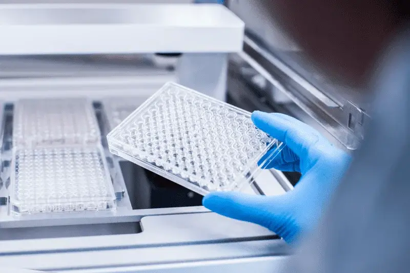
CELLCYTE-X-Optimized-workflows_new
  • Easy setup for user-intuitive experiments
  • Prepare up to 6 vessels concurrently
  • Compatible with multiple vessel types and user protocols
  • Automatically capture images at user-denned time points
  • Acquire images from inside the incubator
  • Maintain optimal environment and maximize cell viability
  • Guide users to conclusive observations and results
  • Produce publication-quality graphs
Screenshot 2026-07-01 at 2.24.57 PM
  • Analyze spheroids
  • Capture images and movies in real-time
  • Flexible scheduling options
  • Automated graphing tools
  • Platemap feature to plan out your studies ahead of time
  • 4x imaging
CELLCYTE-X-Real-time-analysis
  • Streamlined workflow to monitor spheroid formations inside your incubator
  • Ensured autofocus at every timepoint for long-term live cell studies
  • Powerful analysis algorithm for accurate spheroid masking and tracking
  • Diverse analysis metrics supporting a wide range of spheroid assays
  • Maximize throughput and reproducibility by running up to 6 plates in a 96-well plate format
CELLCYTE-X-Optimized-workflows_new
  • Easy setup for user-intuitive experiments
  • Prepare up to 6 vessels concurrently
  • Compatible with multiple vessel types and user protocols
  • Automatically capture images at user-denned time points
  • Acquire images from inside the incubator
  • Maintain optimal environment and maximize cell viability
  • Guide users to conclusive observations and results
  • Produce publication-quality graphs
SPEAK TO OUR EXPERTS

Get a Quote or Book a Demo

GET A QUOTE BOOK A DEMO
UPSIGHT People In Lab